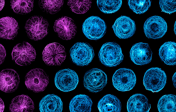
Cellular mechanical effects are classically achieved by the contraction of actin filaments thanks to molecular motors, the myosins. However, actin alone does not allow cells to pass through such small interstices. Researchers at IRIG, in collaboration with teams from CNRS and Utrecht University in Holland, have revealed the vital role played by microtubules in achieving such a morphological transformation. (figure).
Since 2013, as they have developed their studies on the shape and division of stem cells, researchers have discovered how sensitive microtubules are to the pressure forces experienced by the cell in the interstices. The scientists have cleverly developed a device based on a kind of plastic film in which living cells are grafted: by stretching the film, it becomes possible to apply precise deformations to the cells. Forces are applied with perfectly controlled speed, frequency, direction and pressure-relaxation. In this way, microtubules, which are generally highly dynamic, are stabilized when the cells are compressed into the interstices, enabling them to progress. In addition, the researchers identified the molecular mechanism of microtubule stabilization: proteins associated with the growing ends of microtubules ensure their rapid elongation by repositioning themselves in the deformed zones.
These studies reveal new fundamental properties of microtubules and demonstrate their involvement in cell migration in response to the mechanical and spatial constraints of the environment. These processes could well be involved in tumor invasion or tissue exploration by immune cells. The mechanism identified could lead to the development of a new molecular strategy to control cell migration.

Figure: microtubule network architecture.